What is it?
Melanoma, the most serious type of skin cancer, develops in the cells that produce melanin — the pigment that gives your skin its color. Melanoma can also form in your eyes and, rarely, in internal organs, such as your intestines.
The exact cause of all melanomas isn't clear, but exposure to ultraviolet (UV) radiation from sunlight or tanning lamps and beds increases your risk of developing melanoma. Other factors, such as your genetic makeup, is also likely to play a role.
Limiting your sun exposure and avoiding tanning lamps and beds can help reduce your risk of melanoma. And making sure you know the warning signs of skin cancer can help ensure that cancerous changes are detected and treated before the cancer has a chance to spread. Melanoma can be treated successfully if it is detected early.
Symptoms
Melanomas can develop anywhere on your body, but they most often develop in areas that have had exposure to the sun, such as your back, legs, arms and face. Melanomas can also occur in areas that don't receive much sun exposure, such as the soles of your feet, palms of your hands and on fingernail beds. These hidden melanomas are more common in people with darker skin.
The first melanoma symptoms often are:
- A change in an existing mole
- The development of a new, unusual-looking growth on your skin
Melanoma doesn't always begin as a mole. It can also occur on otherwise normal-appearing skin.
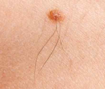
Normal moles
Normal moles are generally a uniform colour, such as tan, brown or black, with a distinct border separating the mole from your surrounding skin. They're oval or round and usually smaller than 1/4 inch (6 millimeters) in diameter — the size of a pencil eraser.
Most people have between 10 and 40 moles. Many of these develop by age 40, although moles may change in appearance over time — some may even disappear with age.
Unusual moles that may indicate melanoma
Characteristics of unusual moles that may indicate melanomas or other skin cancers follow the A-B-C-D-E guide developed by the American Academy of Dermatology:
- A is for asymmetrical shape. Look for moles with irregular shapes, such as two very different-looking halves.
- B is for irregular border. Look for moles with irregular, notched or scalloped borders — characteristics of melanomas.
- C is for changes in colour. Look for growths that have many colours or an uneven distribution of colour.
- D is for diameter. Look for new growth in a mole larger than about 1/4 inch (6 millimeters).
- E is for evolving. Look for changes over time, such as a mole that grows in size or that changes color or shape. Moles may also evolve to develop new signs and symptoms, such as new itchiness or bleeding.
Other suspicious changes in a mole may include:
- Scaliness
- Itching
- Spreading of pigment from the mole into the surrounding skin
- Oozing or bleeding
Cancerous (malignant) moles vary greatly in appearance. Some may show all of the changes listed above, while others may have only one or two unusual characteristics.
Hidden melanomas
Melanomas can also develop in areas of your body that have little or no exposure to the sun, such as the spaces between your toes and on your palms, soles, scalp or genitals. These are sometimes referred to as hidden melanomas, because they occur in places most people wouldn't think to check. When melanoma occurs in people with darker skin, it's more likely to occur in a hidden area.
Hidden melanomas include:
- Melanoma under a nail. Subungual melanoma is a rare form that occurs under a nail and can affect the hands or the feet. It's more common in blacks and in other people with darker skin pigment. The first indication of a subungual melanoma is usually a brown or black discoloration that's often mistaken for a bruise (hematoma).
- Melanoma in the mouth, digestive tract, urinary tract or vagina. Mucosal melanoma develops in the mucous membrane that lines the nose, mouth, esophagus, anus, urinary tract and vagina. Mucosal melanomas are especially difficult to detect because they can easily be mistaken for other, far more common conditions. A melanoma in a woman's vagina can cause itching and bleeding. Anal melanoma can cause anal bleeding and pain during bowel movements. Melanoma that occurs in the esophagus can cause difficulty swallowing.
- Melanoma in the eye. Eye melanoma, also called ocular melanoma, occurs in the uvea — the layer beneath the white of the eye (sclera). An eye melanoma may cause vision changes and may be diagnosed during an eye exam.
Causes
Melanoma occurs when something goes awry in the melanin-producing cells (melanocytes) that give color to your skin. Normally, skin cells develop in a controlled and orderly way — healthy new cells push older cells toward your skin's surface, where they die and eventually are sloughed off. But when some cells develop DNA damage, new cells may begin to grow out of control and can eventually form a mass of cancerous cells.
Just what damages DNA in skin cells and how this leads to melanoma isn't clear. It's likely that a combination of factors, including environmental and genetic factors, causes melanoma. Still, doctors believe exposure to ultraviolet (UV) radiation from the sun and from tanning lamps and beds is the leading cause of melanoma.
UV light doesn't cause all melanomas, especially those that occur in places on your body that don't receive exposure to sunlight. This indicates that other factors may contribute to your risk of melanoma.
Risk factors
Factors that may increase your risk of melanoma include:
- Fair skin. Having less pigment (melanin) in your skin means you have less protection from damaging UV radiation. If you have blonde or red hair, light-colored eyes, and you freckle or sunburn easily, you're more likely to develop melanoma than is someone with a darker complexion. But melanoma can develop in people with darker complexions, including hispanics and blacks.
- A history of sunburn. One or more severe, blistering sunburns as a child or teenager can increase your risk of melanoma as an adult.
- Excessive ultraviolet (UV) light exposure. Exposure to UV radiation, which comes from the sun and from tanning beds, can increase the risk of skin cancer, including melanoma.
- Living closer to the equator or at a higher elevation. People living closer to the earth's equator, where the sun's rays are more direct, experience higher amounts of UV radiation, as compared with those living in higher latitudes. In addition, if you live at a high elevation you're exposed to more UV radiation.
- Having many moles or unusual moles. Having more than 50 ordinary moles on your body indicates an increased risk of melanoma. Also, having an unusual type of mole increases the risk of melanoma. Known medically as dysplastic nevi, these tend to be larger (greater than 1/5 inch or 5 millimeters) than normal moles and have irregular borders and a mixture of colors.
- A family history of melanoma. If a close relative, such as a parent, child or sibling, has had melanoma, you have a greater chance of developing it too.
- Weakened immune system. People with weakened immune systems have an increased risk of skin cancer. This includes people who have HIV/AIDS and those who have undergone organ transplants.
Diagnosis
Skin cancer screening
Ask your doctor whether you should consider periodic screening for skin cancer. You and your doctor may consider screening options such as:
- Skin exams by a trained professional. The American Cancer Society (ACS) recommends periodic skin exams as part of your usual checkups with your doctor. During a skin exam, your doctor conducts a head-to-toe inspection of your skin.
- Skin exams you do at home. In addition, the ACS and the American Academy of Dermatology recommend occasional self-exams. A self-exam may help you learn the moles, freckles and other skin marks that are normal for you, so you can notice any changes. It's best to do this standing in front of a full-length mirror while using a hand-held mirror to inspect hard-to-see areas. Be sure to check the fronts, backs and sides of your arms and legs. In addition, check your groin, scalp, fingernails, your soles and the spaces between your toes.
Other groups don't recommend skin cancer screening exams because it's not clear whether screening saves lives. Instead, finding an unusual mole could lead to a biopsy, which, if the mole is found to not be cancerous, could lead to unnecessary pain, anxiety and cost. Talk to your doctor about what screening is right for you, based on your risk of skin cancer.
Diagnosing melanoma
Sometimes cancer can be detected simply by looking at your skin, but the only way to accurately diagnose melanoma is with a biopsy. In this procedure, all or part of the suspicious mole or growth is removed, and a pathologist analyses the sample. Biopsy procedures used to diagnose melanoma include:
- Punch biopsy. During a punch biopsy, your doctor uses a tool with a circular blade. The blade is pressed into the skin around a suspicious mole and a round piece of skin is removed.
- Excisional biopsy. In this procedure, the entire mole or growth is removed, along with a small border of normal-appearing skin.
- Incisional biopsy. With an incisional biopsy, only the most irregular part of a mole or growth is taken for laboratory analysis.
The type of skin biopsy procedure you undergo will depend on your situation.
Melanoma stages
If you receive a diagnosis of melanoma, the next step is to determine the extent, or stage, of the cancer. To assign a stage to your melanoma, your doctor will:
- Determine the thickness. The thickness of a melanoma is determined by carefully examining the melanoma under a microscope. The thickness of a melanoma helps doctors decide on a treatment plan. In general, the thicker the tumour, the more serious the disease.
- See if the melanoma has spread. To determine whether your melanoma has spread to nearby lymph nodes, your surgeon may use a procedure known as a sentinel node biopsy. During a sentinel node biopsy, a dye is injected in the area where your melanoma was removed. The dye flows to the nearby lymph nodes. The first lymph nodes to take up the dye are removed and tested for cancer cells. If these first lymph nodes (sentinel lymph nodes) are cancer-free, there's a good chance that the melanoma has not spread beyond the area where it was first discovered.
Melanoma is staged using the Roman numerals I through IV. A stage I melanoma is small and has a very successful treatment rate. But the higher the numeral, the lower the chances of a full recovery. By stage IV, the cancer has spread beyond your skin to other organs, such as your lungs or liver.
References
http://www.cancer.ie/cancer-information/melanoma#sthash.z6FuijC7.dpbs
http://emedicine.medscape.com/article/280245-overview
http://www.nhs.uk/conditions/malignant-melanoma/Pages/Introduction.aspx
http://www.skincancer.org/skin-cancer-information/melanoma/the-stages-of-melanoma/guide-to-staging-melanoma