What is it?
Pleurisy occurs when the double membrane (pleura) that lines your chest cavity and surrounds each of your lungs becomes inflamed. Also called pleuritis, pleurisy typically causes sharp pain, almost always when you take a breath.
Pleurisy occurs as a complication of a wide variety of underlying conditions. Relieving pleurisy involves treating the underlying condition, if it's known, and taking pain relievers.
Symptoms
The signs and symptoms of pleurisy may include:
- Chest pain when you inhale and exhale (between breaths, you feel almost no pain)
- Shortness of breath
- Dry cough
- Fever and chills, depending on the cause
The sharp, fleeting pain in your chest that pleurisy causes is made worse by coughing, sneezing, moving and deep breathing. In some cases, pain may extend from your chest to your shoulder. You may find relief from pain when you hold your breath or when you apply pressure over the painful area.
When an accumulation of fluids (pleural effusion) is associated with pleurisy, the pain usually disappears because the fluid serves as a lubricant. However, if enough fluid accumulates, it puts pressure on your lungs and interferes with their normal function, causing shortness of breath. If the fluid becomes infected, the signs and symptoms of dry cough, fever and chills may appear. An infected pleural effusion is called an empyema.
Causes
A double layer of membranes called pleura separate your lungs from your chest wall. One layer of the pleura overlies each lung. The other layer lines the inner chest wall. The layers are like two pieces of smooth satin rubbing against each other with almost no friction, allowing your lungs to expand and contract when you breathe without any resistance from the lining of the chest wall.
When inflamed, the two layers of the pleural membrane in the affected side of your chest rub against each other like two pieces of sandpaper, producing the pain of pleurisy when you inhale and exhale.
The underlying medical conditions that can cause pleurisy are numerous. Pleurisy causes include:
- An acute viral infection, such as the flu (influenza)
- Pneumonia, in those cases in which the infected portion of your lung involves the surface of the pleura
- Autoimmune conditions, such as lupus, rheumatoid arthritis and autoimmune hepatitis
- Tuberculosis and other infections
- A clot in an artery of your lungs (pulmonary embolism)
Pleurisy can also occur as a result of trauma to your chest or after heart surgery. Rib fractures also may cause pleurisy. It's possible to fracture a rib in the absence of trauma, such as from a severe cough. In some cases, the cause of pleurisy is unknown (idiopathic).
Cancer in the lung rarely causes pleurisy. There's no relationship between smoking and pleurisy, but a "smoker's cough" will aggravate the pain of this condition.
Diagnosis
Your doctor may make a diagnosis based on your signs and symptoms. When examining you and listening to your chest, your doctor may hear a "friction rub" that may sound like the crunching sound of walking on very dry snow.
Your doctor might also use the following procedures to determine the underlying cause of pleurisy:
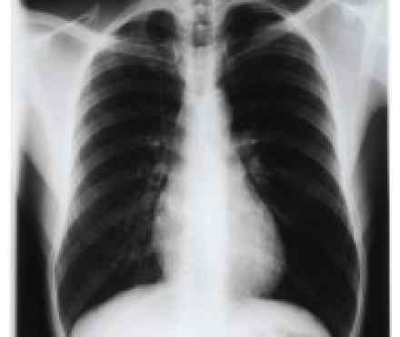
- Imaging. A chest X-ray may show an area of inflammation in your lungs that indicates pneumonia. Your doctor will want to investigate an unexplained abnormality seen on an X-ray with additional imaging, usually beginning with a computerized tomography (CT) scan. In a CT procedure, a computer translates information from X-rays into images of thin sections (slices) of your chest. CT scans produce more-detailed images of your internal organs than do conventional X-ray studies. Sometimes doctors want a special type of chest X-ray in which you lie on your side where the pleurisy is to see if there's any fluid that doesn't appear on a standard chest X-ray. This type of X-ray is called a decubitus chest X-ray. Your doctor may also use ultrasound to determine whether you have a pleural effusion.
- Blood test. A blood test may tell your doctor if you have an infection and, if so, what type of infection you have. Other blood tests also may detect an autoimmune disorder, such as rheumatoid arthritis or lupus, in which the initial sign is pleurisy.
- Thoracentesis. To remove fluid for laboratory analysis, your doctor may suggest a procedure called thoracentesis. In this procedure, your doctor first injects a local anesthetic, then inserts a needle through your chest wall between your ribs to remove fluid. In addition, a sample of tissue (pleural biopsy) for microscopic analysis may be obtained if your doctor is concerned that the fluid collection may be caused by tuberculosis or cancer. If only a small amount of fluid is present, your doctor may insert the needle with the help of ultrasound guidance over the site of the fluid.
- Thoracoscopy. This procedure allows a surgeon to see inside your chest and obtain a sample of pleural tissue. First, the surgeon makes one or more small incisions between your ribs. A tube with a tiny video camera is then inserted into your chest cavity — a procedure sometimes called video-assisted thoracoscopic surgery (VATS). Special surgical tools allow your surgeon to cut away tissue for testing.
References
http://www.medicinenet.com/pleurisy/article.htm
http://www.nhs.uk/Conditions/Pleurisy/Pages/Introduction.aspx
http://www.healthline.com/health/pleurisy
https://www.hse.ie/eng/health/az/P/Pleurisy/Causes-of-pleurisy.html